File:Ct-workstation-neck.jpg
Original file (1,026 × 1,026 pixels, file size: 225 KB, MIME type: image/jpeg)
| This free media file is from Wikimedia Commons. Its description page is included below. |
| Description |
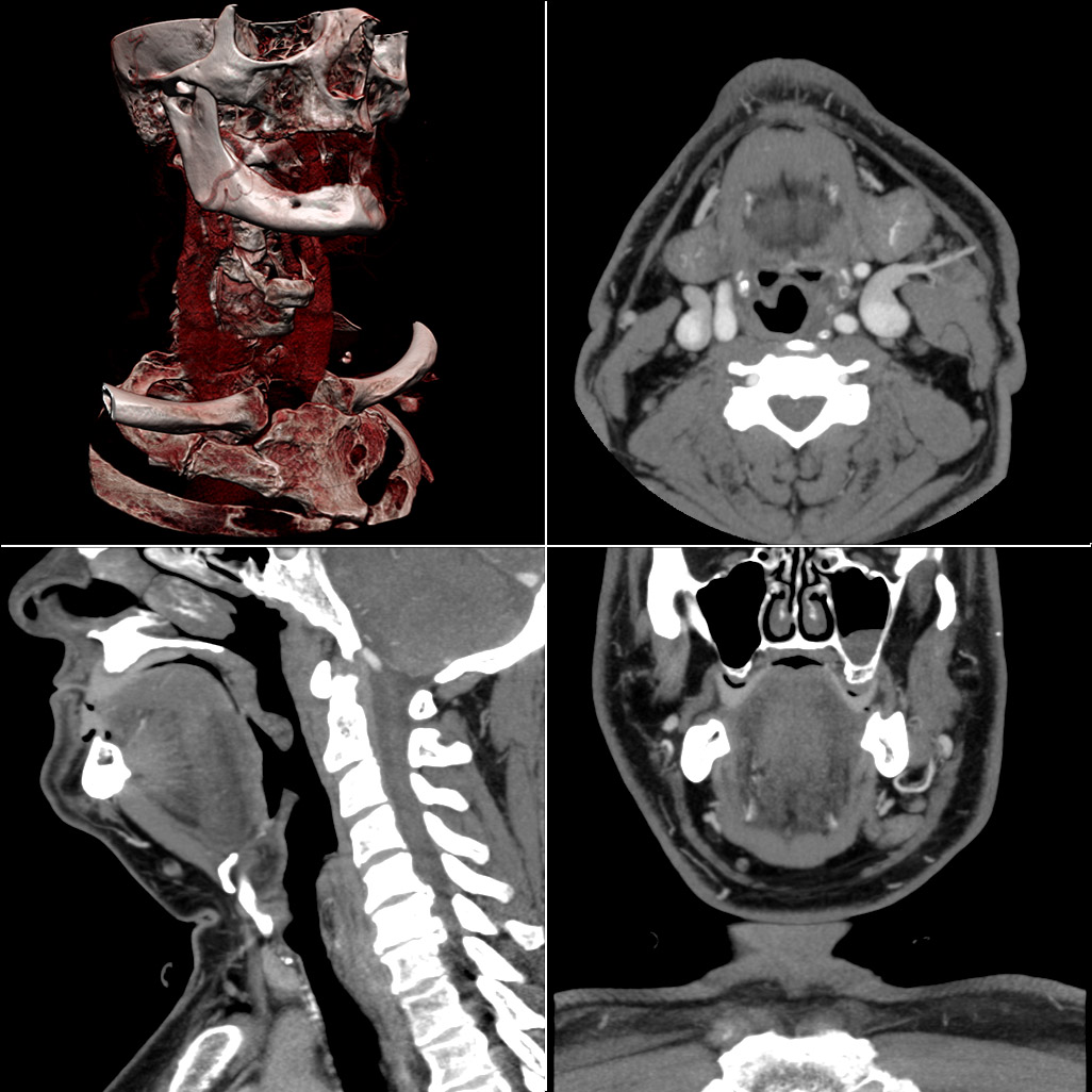
Typical screen layout of workstation software used for reviewing multi-detector CT studies. Clockwise from top-left: Volume rendering overview, axial slices, coronal slices, sagittal slices. A study may consist of several hundred slices which the user can scroll through. Images are usually acquired by the scanner in the 'axial' plane. The workstation reconstructs coronal, sagittal or oblique images on demand. Although visually very appealing, the volume rendering is often of limited diagnostic value, and requires substantial computer resources. Qualitative and quantitative information tends to be more accessible on the cross-sectional images, and many operators prefer to forgo the volume rendering for an oblique cross-sectional series, or a duplicate series displayed with different window settings. Sophisticated workstation software may include curved-plane cross-sectional reconstructions (which is able to 'straighten' a meandering blood vessel so that accurate measurements can be made), and image segmentation tools (e.g. for semi-automatic calculation of coronary artery calcium content). |
||||||||
| Date | |||||||||
| Source | http://en.wikipedia.org/wiki/File:Ct-workstation-neck.jpg | ||||||||
| Author | en:User:ChumpusRex | ||||||||
| Permission (Reusing this file) |
I, the copyright holder of this work, hereby publish it under the following license:
|

File history
Click on a date/time to view the file as it appeared at that time.
| Date/Time | Thumbnail | Dimensions | User | Comment | |
|---|---|---|---|---|---|
| current | 16:27, 23 February 2009 | | 1,026 × 1,026 (225 KB) | Linforest | {{Information |Description=Typical screen layout of workstation software used for reviewing multi-detector CT studies. Clockwise from top-left: Volume rendering overview, axial slices, coronal slices, sagittal slices. A study may consist of several hund |
File usage
The following page uses this file:
Global file usage
The following other wikis use this file:
- Usage on ar.wikipedia.org
- Usage on as.wikipedia.org
- Usage on bs.wikipedia.org
- Usage on ca.wikipedia.org
- Usage on de.wikipedia.org
- Usage on en.wikipedia.org
- Usage on en.wikiversity.org
- Usage on es.wikipedia.org
- Usage on fr.wikipedia.org
- Usage on fr.wiktionary.org
- Usage on hr.wikipedia.org
- Usage on it.wikipedia.org
- Usage on ja.wikipedia.org
- Usage on ml.wikipedia.org
- Usage on nl.wiktionary.org
- Usage on ru.wikipedia.org
- Usage on sh.wikipedia.org
- Usage on sr.wikipedia.org
- Usage on zh.wikipedia.org